Hemisphereectomy And Postoperative Time
Hemisphereectomy is a surgical procedure used to treat a number of seizure-causing diseases. It is typically used when other, less radical therapies are not effective in the disease.
The first hemisphereectomy was performed on a dog in 1888. The first reference to performing the procedure on humans dates back to 1923. In the 60s and 70s, several procedures were performed with poor results.
Today, things are different. Physicians choose anatomical hemisphereectomy over functional hemisphereectomy. It is a more accurate and less invasive procedure with a much higher probability of success than in the past.
In this article, we will take a closer look at what this method is like today.
What is a hemisphereectomy?
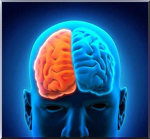
Hemisphereectomy is a neurosurgical procedure in which one of the hemispheres of the brain is removed. Sometimes the surgeon removes the left hemisphere, sometimes the right hemisphere. This procedure is performed only in extreme cases and only for children aged 5-10 years.
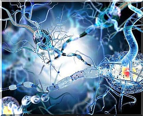
This type of procedure is done mainly to prevent epileptic seizures. It is also useful in patients with neurological deficits or severe head injury.
Most procedures remove the entire other half of the brain, but sometimes only part of it. The latter is called a functional hemisphereectomy. In these cases, if doctors leave a small portion of the damaged tissue behind, the seizures may recur.
Hemispherectomy: treatment issues
In general, hemispherectomy may be used in patients with persistent and daily epileptic seizures that are not responding to drug therapy or other less invasive surgical procedures.
Hemispherectomy is recommended in the following cases:
- For children with hemiplegia. This can only be done in children over four years of age who suffer from epileptic seizures and / or mental disorders and after trying drug treatment for two years without success.
- Sturge-Weber syndrome. It is a nerve-skin disease characterized by the birth of a face in the area of the trigeminal nerve. Doctors may recommend this type of surgery when epileptic seizures begin at an early age and the disease affects the entire hemisphere.
- Rasmussen’s encephalitis. This brain disease causes chronic, progressive encephalitis, or encephalitis. Early treatment is important.
- Hemimegalencephaly. This is a rare inflammatory neurological disease that causes severe epileptic seizures. Doctors still disagree on whether surgery is the best option for patients with this disease.
- Deformities in cortical development.
What kind of hemisphereectomy is the procedure?
There are four different types of hemispherectomy, described below.
- Anatomical hemisphereectomy
- Brain decortication
- Functional hemisphereectomy
- Modified functional hemisphereectomy
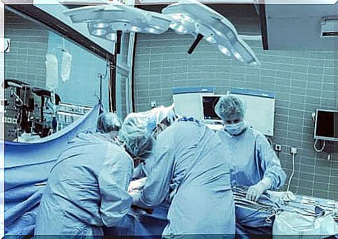
Usually the procedure uses general anesthesia. The surgeon begins by shaving the patient’s head and marking the lines of surgery. The surgeon then cuts to reveal the sclera. It is removed to make the brain visible.
The area to be deleted is then carefully marked. The area is cut, and after the cut, the blood vessels are burned. A drainage tube is placed in the brain. The sclera and scalp are put back on and the incision is closed with rivets.
Postoperative time
The postoperative time is very painful. Usually the drain pipe is left in place for 3-4 days. The doctor then makes an assessment of the patient and decides whether or not to remove the tube. Prior to its removal, some diagnostic tests should be performed to determine possible bleeding.
The major complications are hemodynamic instability, hypothermia, and hypo- or hyperkalemia. In general, these problems can be effectively controlled.
Seizures immediately after surgery are another serious complication. Typically, about half of patients develop hydrocephalus, and almost all patients develop aseptic meningitis. Nevertheless, the mortality rate is quite low and ranges from 4 to 6%. There is also evidence that some of the complications may not appear until later.
Seizures resolve in 70-85% of patients who have undergone a hemisphereectomy. By about 10-20%, the quality of life improves significantly.